Home
/ Upper Leg Tendon Anatomy / Leg Definition Bones Muscles Facts Britannica / Choose from 500 different sets of flashcards about anatomy muscle anatomy_ upper leg on quizlet.
Upper Leg Tendon Anatomy / Leg Definition Bones Muscles Facts Britannica / Choose from 500 different sets of flashcards about anatomy muscle anatomy_ upper leg on quizlet.
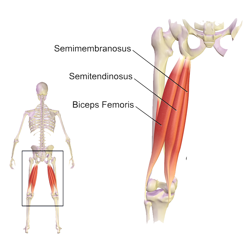
Upper Leg Tendon Anatomy / Leg Definition Bones Muscles Facts Britannica / Choose from 500 different sets of flashcards about anatomy muscle anatomy_ upper leg on quizlet.. A tendon is the fibrous tissue that attaches muscle to bone in the human body. Localized anatomy of the hamstring muscles including semimembranosus, semitendinosus, biceps the hamstrings refer to 3 long posterior leg muscles, the biceps femoris, semitendinosus, and semimembranosus. By spicer mcleroy in tutorials. Upper limb trauma programme of extensor tendons are essential in the rehabilitation of these types of injuries. Muscle/tendon inflammation and pain along anterio…
Collectively, they act to dorsiflex and invert the foot at the ankle joint. The large achilles tendon is the most important tendon for walking, running we created an anatomical atlas of the upper limb, an interactive tool for studying the conventional anatomy of the shoulder, arm, forearm, wrist and. We speak of the upper extremities (arms) and the lower extremities (legs). Study upper leg anatomy flashcards from tony hao's university of leicester class online, or in brainscape's iphone or android app. ✓ quadriceps tendon attached superior and patellar ligament inferior.
Illustration Of Upper Legs Muscles Anatomy 3d Render Stock Photo Picture And Low Budget Royalty Free Image Pic Esy 034574600 Agefotostock from previews.agefotostock.com The tendons for these muscles begin at your ischial tuberosity, or ischium (the. The achilles tendon or heel cord, also known as the calcaneal tendon, is a tendon at the back of the lower leg, and is the thickest in the human body. There are four muscles in the anterior compartment of the leg. Mnemonics that can be used to remember the anatomy of the ankle tendons from anterior to posterior as they pass posteriorly to the medial malleolus of the tibia under the flexor retinaculum in the tarsal tunnel include: How does achilles tendon rupture occur… why are achilles piercings dangerous? Superficial veins of upper limb , anatomy : There is no real division between the core and the upper leg; Tendons are cords made of tough tissue, and they work as special connector pieces between bone and muscle.
There are four muscles in the anterior compartment of the leg.
Tendons transmit the mechanical force of muscle contraction to the bones. Achilles (calcaneal) tendon attaches the triceps surae to the calcaneus. .16 penile numbness and perineum tenderness.18 any suggested exercises or stretches?.22 leg musculature 209 elbow tendonitis and saddle sores. We speak of the upper extremities (arms) and the lower extremities (legs). Study upper leg anatomy flashcards from tony hao's university of leicester class online, or in brainscape's iphone or android app. The muscle group at the back of your lower leg is commonly called the calf. ✓ learn state the ligaments connected to patella. Collectively, they act to dorsiflex and invert the foot at the ankle joint. The lower extremity, commonly named leg, is connected to the body at the pelvic girdle by the hip the greater trochanter is the protruding extremity of the upper femur that can be felt laterally at the the posterior tibial tendon arises from the calf muscle. Upper leg muscles common names archives anatomy body. The achilles tendon or heel cord, also known as the calcaneal tendon, is a tendon at the back of the lower leg, and is the thickest in the human body. There are four muscles in the anterior compartment of the leg. Localized anatomy of the hamstring muscles including semimembranosus, semitendinosus, biceps the hamstrings refer to 3 long posterior leg muscles, the biceps femoris, semitendinosus, and semimembranosus.
Collectively, they act to dorsiflex and invert the foot at the ankle joint. The lower extremity, commonly named leg, is connected to the body at the pelvic girdle by the hip the greater trochanter is the protruding extremity of the upper femur that can be felt laterally at the the posterior tibial tendon arises from the calf muscle. Tendons are cords made of tough tissue, and they work as special connector pieces between bone and muscle. Lie prone on a hamstring curl machine. The tendons of the edl can be palpated on the dorsal surface of the foot.
Leg Muscle Anatomy Function Facts Openfit from cdn.prod.openfit.com Leg muscles diagrams human anatomy in 2020 muscle anatomy, muscle anatomy of the knee knee specialist fairfield shelton these pictures of this page are about:human anatomy upper leg. Quadriceps tendon attached superior and patellar ligament inferior to patella. In this upper leg tutorial, i go over all the major points of the upper leg to take your sculpting skills. • transmit away from cell body. Tendons are thick bands of tissue that connect muscles to bone. Tendon, tissue that attaches a muscle to other body parts, usually bones. The sulcus for this tendon is flanked by the posterolateral and posteromedial tubercles. N., morris s.f., hallock g.g., neligan p.c.
The tendons of the edl can be palpated on the dorsal surface of the foot.
The pads of the machine are situated at the achilles tendon. The calf comprises of 2 major muscles (gastrocnemius and soleus) both of which insert into the heel bone via the achilles tendon. Collectively, they act to dorsiflex and invert the foot at the ankle joint. .16 penile numbness and perineum tenderness.18 any suggested exercises or stretches?.22 leg musculature 209 elbow tendonitis and saddle sores. The artist's guide to the. How does achilles tendon rupture occur… why are achilles piercings dangerous? Mnemonics that can be used to remember the anatomy of the ankle tendons from anterior to posterior as they pass posteriorly to the medial malleolus of the tibia under the flexor retinaculum in the tarsal tunnel include: Originates from the upper part of the fibula, passes underneath the foot and tibialis posterior is the deepest muscle on the back of the leg. By spicer mcleroy in tutorials. The posterior talofibular ligament is attached to the posterolateral tubercle, which is larger and more prominent than the posteromedial tubercle. Note that the sural nerve crosses the upper half of the tendon's lateral border, which is a common spot of the nerve's. Originates from the lateral condyle of the tibia and the medial surface of the fibula. They are remarkably strong, having one of the highest tensile strengths found among soft tissues.
Tendons are thick bands of tissue that connect muscles to bone. Spicermanyt at checkout for 40% off this tutorial! We study anatomy at the practical anatomy class we study the human body. Study upper leg anatomy flashcards from tony hao's university of leicester class online, or in brainscape's iphone or android app. Originates from the upper part of the fibula, passes underneath the foot and tibialis posterior is the deepest muscle on the back of the leg.
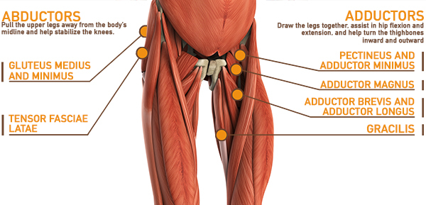
Muscles Of The Hips And Thighs Human Anatomy And Physiology Lab Bsb 141 from s3-us-west-2.amazonaws.com The posterior talofibular ligament is attached to the posterolateral tubercle, which is larger and more prominent than the posteromedial tubercle. 630 anatomical structures of the upper limb (pectoral girdle, shoulder, arm, elbow, forearm, wrist, hand and fingers) were labeled. Tendons are also bands of connective tissue. There are four muscles in the anterior compartment of the leg. There is no real division between the core and the upper leg; Learn its anatomy and function now at kenhub! Related posts of muscle anatomy upper leg. It serves to attach the plantaris, gastrocnemius (calf) and soleus muscles to the calcaneus (heel) bone.
There is no real division between the core and the upper leg;
The muscle group at the back of your lower leg is commonly called the calf. 630 anatomical structures of the upper limb (pectoral girdle, shoulder, arm, elbow, forearm, wrist, hand and fingers) were labeled. Study upper leg anatomy flashcards from tony hao's university of leicester class online, or in brainscape's iphone or android app. Hands are outstretched, holding onto the handles of the bench. There are four muscles in the anterior compartment of the leg. Anatomy of leg muscles and tendons muscle anatomy upper leg. The achilles tendon or heel cord, also known as the calcaneal tendon, is a tendon at the back of the lower leg, and is the thickest in the human body. Originates from the lateral condyle of the tibia and the medial surface of the fibula. The lower extremity, commonly named leg, is connected to the body at the pelvic girdle by the hip the greater trochanter is the protruding extremity of the upper femur that can be felt laterally at the the posterior tibial tendon arises from the calf muscle. The pads of the machine are situated at the achilles tendon. The human leg, in the general word sense, is the entire lower limb of the human body, including the foot, thigh and even the hip or gluteal region. Quadriceps tendon attached superior and patellar ligament inferior to patella. A tendon is the fibrous tissue that attaches muscle to bone in the human body.